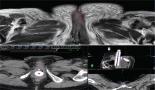
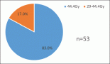
With the purpose of implementing a way to obtain volumes from ultrasound axial images, a novel method for 3D gynaecologic brachytherapy was assessed, with a 3D-printed attachment for a commercial stepper (for prostate brachytherapy). It allowed the acquisition of a transabdominal axial image series by ultrasound; these images were uploaded to a treatment planning system where high-risk clinical tumour volume (HR-CTV) and risk organs were contoured. A dose administration plan was developed with orthogonal X-ray images (0° and 270° incidences), using International Commission on Radiation Units and Measurements (ICRU) 38 points. The same plan was applied in the ultrasound images’ sequence and their respective volumes; differences were noted. In the 20 cases analysed, with a given point A dose, its corresponding dose delivered to 90% of HR-CTV percentage was highly variable (mean = 104.2, SD = 26.01). There is a significant variation of this percentage when point A falls outside the HR-CTV than when it falls inside (p < 0.00001). There is a significant correlation in terms of the bladder point dose ICRU 38 and the Maximum dose to 2cc of organ or target volume (D2cc) bladder (p = 0.021); however, there is no such correlation when we relate the rectum point dose ICRU 38 to the D2cc rectum (p = 0.327). There was a negative correlation between HR-CTV and D2cc bladder and D2cc rectum; both were statistically significant. There were significant differences comparing ICRU points and dose to prescription and organ at risk volumes, pointing out that there is room for optimisation of plans using the latter technique. So, it is proposed to further test this image modality and compare it to other imaging techniques that allow the creation of volumes, such as computed tomography or magnetic resonance imaging.
Supporting oncology professionals through education
The content on this site is intended for healthcare professionals only