Primary renal lymphoma—a case report
N Geetha1, Abdul Shahid1, Varun Rajan1 and Priya Mary Jacob2
1Department of Medical Oncology, Regional Cancer Centre, Trivandrum 695011, India
2Department of Pathology, Regional Cancer Centre, Trivandrum 695011, India
Correspondence to: Geetha N. Email: geenarayanan@yahoo.com
Abstract
Primary renal lymphoma is a rare entity representing less than 1% of lesions in the kidney. We present the case of a 42-year-old male who was evaluated for pain and a mass in the abdomen. The computed tomogram of the abdomen showed a large lobulated homogeneously enhancing mass lesion of about 14×12×18 cm, involving the whole of the left kidney and encasing the left renal vessels and ureter. The patient underwent a biopsy, and the histopathology was diffuse large B cell lymphoma, positive for LCA, CD20, PAX 5, Bcl 2 and negative for SIgM, CD33, CD34, CD5, Tdt, with MIB 1 labelling index of 40%. He received chemotherapy with rituximab, cyclophosphamide, doxorubicin, vincristine and prednisolone (R CHOP) for eight cycles followed by radiation to the residual mass and achieved complete remission. Currently, he is alive and in remission at 28 months.
Keywords: NHL, Kidney
Copyright: © the authors; licensee ecancermedicalscience. This is an Open Access article distributed under the terms of the Creative Commons Attribution License (http://creativecommons.org/licenses/by/3.0), which permits unrestricted use, distribution, and reproduction in any medium, provided the original work is properly cited.
Introduction
Primary renal lymphoma (PRL) is a rare entity of the genitourinary tract in which the disease is localized in the kidneys or in which renal involvement is the presenting feature [1]. Renal involvement is detected in 3–8% of all patients undergoing staging for non-Hodgkin’s lymphoma (NHL); however, in autopsy series, renal involvement has been reported in 30–60% [2]. PRL represents less than 1% of the lesions in this organ [3]. Here, we report on a patient with PRL.
Case Report
A 42-year-old male was evaluated for abdominal pain and a mass in the abdomen at the local hospital. The examination showed a firm lobulated mass on the left side of the abdomen. The ultrasonogram showed a large renal mass on the left side, crossing the midline anterior to the aorta, extending to the epigastrium, left iliac region and involving the tail of the pancreas. The computed tomogram (CT) of the abdomen with urographic sequence showed a large lobulated homogeneously enhancing mass lesion of about 14×12×18 cm, involving the whole of the left kidney and encasing the left renal vessels and ureter (Figures 1 and 2). The mass was seen to be displacing the bowel loops to the right side and the splenic vessels anteriorly. On urographic sequence, the left kidney did not show any excretion (Figure 3). Multiple enlarged lymphnodes in the paraaortic, retrocaval, renal hilar, mesenteric region of 10–40 mm in size were present.
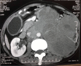
Figure 1. A contrast-enhanced CT of the abdomen showing large, lobulated homogenously enhancing mass lesion 14×12×18 cm arising from left kidney and encasing the renal vessels.
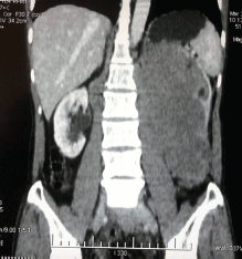
Figure 2. A coronal view showing the mass encasing the left ureter, left renal artery, and vein. The right kidney appears normal.
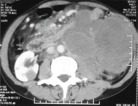
Figure 3. A urographic sequence of the left kidney does not show any excretion.
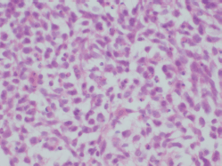
Figure 4. H & E × 400: Tumour cells have moderate cytoplasm, large vesicular nuclei.
The patient underwent biopsy of the mass and subsequently presented to us. He did not have B symptoms. There were no palpable lymphnodes or hepatosplenomegaly or any other mass. His haemogram was normal, and serum chemistries were normal except for a serum creatinine of 1.7 mg/dl and LDH of 2320 U/dl. The histopathological examination of the biopsy showed predominantly large cells with moderate cytoplasm, large vesicular nuclei, prominent nucleoli (Figure 4). Immunohistochemistry analysis showed that the tumour cells were positive for LCA, CD20, PAX 5, Bcl 2 and negative for SIgM, CD33, CD34, CD5, Tdt and MIB 1 labelling index was 40% (Figures 5 and 6). The diagnosis was NHL diffuse large B cell type (DLBCL).
The CT of chest and the biopsy of bone marrow were normal. He received chemotherapy with rituximab, cyclophosphamide, vincristine, doxorubicin, and prednisolone for eight cycles (RCHOP). A positron emission tomogram at the end of chemotherapy showed the perinephric region to be FDG avid, and he was consolidated with irradiation of 36GY/18#. A repeat CT showed no evidence of disease. He is alive in complete remission at 28 months.
Discussion
PRL is defined as lymphoma arising in the renal parenchyma and not resulting from the invasion of an adjacent lymphomatous mass [4]. Secondary involvement of the kidneys by lymphoma usually occurs in disseminated cases. PRL comprises only 0.7% of extranodal lymphomas [5]. Renal involvement is more common in the NHL, and mostly, they are intermediate or high grade B cell lymphomas [5]. PRL is a rare and debated entity since the kidneys are devoid of lymphatic tissue [6]. The proposed pathogenetic mechanisms include origin in the lymphatics of renal capsule or subcapsular lymphatic tissue, with subsequent invasion of the renal parenchyma or any chronic inflammatory process might be the cause of lymphomas [7].
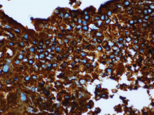
Figure 5. IHC, CD 20 × 400: Tumour cells are CD 20 positive.
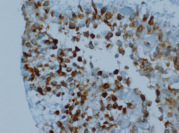
Figure 6. IHC, Bcl 2 × 400: Tumour cells are Bcl 2 positive.
It usually occurs in the middle aged, with the presenting symptoms being flank pain and abdominal mass. The laboratory values are usually normal except for an elevated serum creatinine [4]. Renal lymphoma may present as a solitary mass (10–20%) or multiple mass (60%), and extend by contiguity (25–30%), diffuse infiltration (20%) or perirenal involvement (10%) [8]. Our patient also had a similar presentation.
Yasunaga et al reported eight cases of PRL, six being DLBCL, and none had lymphadenopathy or hepatosplenomegaly at presentation. Their mean survival was six months. In autopsy, only one was limited to the kidney, and the others had extrarenal visceral invasion [7]. A patient with a solitary renal mass invading Gerota’s fascia, with lymph nodes in the mediastinum and retroperitoneum was reported by Pinggera et al, and this case was accepted as possible PRL [6]. Three cases with solitary mass, each one with paraaortic, retrocaval conglomerated lymph nodes and invasion of renal vein and perirenal adipose tissue were described [9]. All cases were accepted as PRL, NHL DLBCL. A 62-year-old male with PRL with lung nodules who had a nephrectomy done and histology was DLBCL [2]. Our patient also had NHL DLBCL positive for LCA, CD 20, PAX5 and BCL 2. A 23-year-old woman with primary renal DLBCL received RCHOP and is alive at 15 months [10]. Pahwa reported on a 39-year-old male with left PRL with local invasion who underwent radical nephrectomy. He was treated with CHOP and was alive at two years [11]. Vedovo et al described an 82-year-old female who underwent radical nephrectomy for a recurrent renal mass and was diagnosed as lymphoplasmacytic MALT lymphoma [12].
In short, there has been no concordance between researchers. Some studies empahsise the absence of lymph node involvement and leukaemic blood picture to make a PRL diagnosis, but in other studies, concurrent lymph node involvement alongside a solitary renal mass, is accepted PRL [7]. Our patient also had a few small paraaortic lymph nodes in addition to the huge renal mass.
It is a diagnostic challenge to differentiate renal lymphoma from other renal masses, especially in cases of unilateral lesions since they simulate renal carcinomas both radiologically and pathologically. Renal lesions that lack typical radiological features of renal cell carcinoma or multi-nodular involvement of the kidney along with lymphadenopathy merits a biopsy. A solitary unilateral renal mass, perirenal mass with distortion of the renal architecture in the CT and the absence of lymph node enlargements are more suggestive of renal cell carcinomas [13]. The standard management of a renal mass is nephrectomy. However, primary renal lymphoma is an exception in which patient can be treated with systemic chemotherapy. Therefore, in spite of its uncommon occurrence, it is of immense importance to distinguish primary renal lymphoma from renal carcinoma.
In view of its aggressive nature and poor prognosis, it is important to make an early diagnosis and start treatment promptly. The treatment of choice is systemic chemotherapy using a CHOP regimen. The earlier reviews report a poor prognosis for patients with PRL, with a median survival of less than one year [4, 7], but the recent reports suggest a better survival probably due to the addition of rituximab to the combination chemotherapy [9, 11]. The present case also received RCHOP and is alive in remission after two years.
Although PRL is a rare tumour type, it must be taken into account when making a differential diagnosis of any renal mass. A high index of suspicion for PRL should be kept if the lesion shows multi-nodular involvement of the kidney along with lymphadenopathy. An effort should be made to make a preoperative diagnosis since primary renal lymphoma can be treated with systemic chemotherapy unlike other renal tumours where one requires nephrectomy.
Conflicts of interest
The authors have no conflicts of interest to declare.
References
1. Tefekli A et al (2006) Lymphoma of the kidney: primary or initial manifestation of rapidly progressive systemic disease? Int Urol Nephrol 38 775–8 DOI: 10.1007/s11255-005-4030-7 PMID: 17111087
2. Ladha A and Haider G (2008) Primary renal lymphoma J Coll Physicians Surg Pak 18 584–5 PMID: 18803901
3. Stallone G et al (2000) Primary renal lymphoma does exist: case report and review of the literature J Nephrol 13 367–72 PMID: 11063141
4. Okuno SH et al (1995) Primary renal non-Hodgkin’s lymphoma – an unusual extranodal site Cancer 75 2258–61 DOI: 10.1002/1097-0142(19950501)75:9<2258::AID-CNCR2820750911>3.0.CO;2-S PMID: 7712433
5. Qiu L et al (2006) Low-grade mucosa-associated lymphoid tissue lymphoma involving the kidney: report of 3 cases and review of the literature Arch Pathol Lab Med 130 86–9 PMID: 16390244
6. Pinggera GM et al (2009) A possible case of primary renal lymphoma: a case report Cases J 2 6233 DOI: 10.4076/1757-1626-2-6233 PMID: 19829773 PMCID: 2740007
7. Yasunaga Y et al (1997) Malignant lymphoma of the kidney J Surg Oncol 64 207–11 DOI: 10.1002/(SICI)1096-9098(199703)64:3<207::AID-JSO6>3.0.CO;2-E PMID: 9121151
8. Urban BA and Fishman EK (2000) Renal lymphoma: CT patterns with emphasis on helical CT Radiographics 20 197–212 DOI: 10.1148/radiographics.20.1.g00ja09197 PMID: 10682781
9. Vázquez Alonso F et al (2009) Primary renal lymphoma: report of three new cases and literature review Arch Esp Urol 62 461–5 PMID: 19736375
10. El fatemi H et al (2012) Uncommon presentation of non-Hodgkin’s lymphoma: a case report and literature review JPBMS 21(6) 1–2
11. Pahwa M et al (2013) Primary renal lymphoma: Is prognosis really that bad? Saudi J Kidney Dis Transpl 24 816–7 DOI: 10.4103/1319-2442.113905 PMID: 23816741
12. Vedovo F et al (2014) Primary renal MALToma: a rare differential diagnosis for a recurrent renal mass after primary ablative therapy Can Urol Assoc J 8(5–6) DOI: 10.5489/cuaj.1645 PMID: 25024802 PMCID: 4081263
13. Dimopoulos MA et al (1996) Primary renal lymphoma: a clinical and radiological study J Urol 155 1865–7 DOI: 10.1016/S0022-5347(01)66031-2 PMID: 8618275


