Oral verruciform xanthoma (OVX) of lower labial mucosa, presenting as a plaque in tobacco users: an unusual case report with literature review
Sandhya Tamgadge1, Treville Pereira1, Aditi Vaidya1 and Vishal Punjabi2
1Department of Oral & Maxillofacial Pathology and Microbiology, D.Y. Patil University School of Dentistry, Sector 7, Nerul, Navi Mumbai 400706, Maharashtra, India
2Department of Oral Pathology and Microbiology, D.Y. Patil University School of Dentistry, Sector 7, Nerul, Navi Mumbai 400706, Maharashtra, India
Abstract
Oral verruciform xanthoma (OVX) is an uncommon lesion known for its wart-like appearance, primarily affecting the oral mucosa. This case report delves into a distinctive presentation of OVX, a rare benign lesion typically characterised by its manifestation as a white plaque in the oral cavity. Clinical features, histological findings, pathogenesis and their implications in the context of differential diagnosis has been discussed. The patient had a history of regular tobacco use, and despite initially presenting as a white plaque, the histopathological and immunohistochemical features strongly suggested verruciform xanthoma.
Keywords: oral verruciform xanthoma, lower lip, white plaque, tobacco, CD 68
Correspondence to: Sandhya Tamgadge
Email: sandhya.tamgadge@gmail.com
Published: 13/11/2024
Received: 18/05/2024
Publication costs for this article were supported by ecancer (UK Charity number 1176307).
Copyright: © the authors; licensee ecancermedicalscience. This is an Open Access article distributed under the terms of the Creative Commons Attribution License (http://creativecommons.org/licenses/by/4.0), which permits unrestricted use, distribution, and reproduction in any medium, provided the original work is properly cited.
Introduction
Oral verruciform xanthoma (OVX) is an infrequent, benign lesion of the oral mucosa, first described by Shafer [1] in 1971. Typically, OVX presents as a well-demarcated, elevated and verrucous lesion [2]. However, this case report highlights an atypical presentation as smooth white plaques resembling leukoplakia, creating a diagnostic dilemma.
OVX has been reported in various oral locations, including the gingiva, hard palate, tongue and buccal mucosa [3, 4]. Notably, its occurrence on the lower labial mucosa is extremely rare. Belknap et al [3] reported that only 1.86% of verruciform xanthoma cases occur on the lower lip.
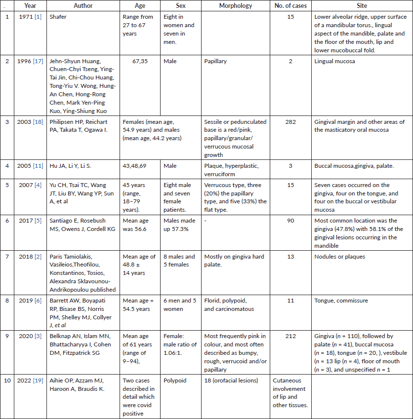
Several retrospective studies have contributed to our understanding of OVX. Santiago et al [5] analysed 90 cases, while Belknap et al [3] presented a substantial series of 212 cases. Yu et al [4] conducted a clinicopathological study of 15 cases, and Tamiolakis et al [2] reported 13 new cases. Barrett et al [6] presented (Table 1) eight typical and three anomalous cases, underscoring the diverse presentations of OVX.
The pathogenesis of OVX remains unclear, with various associated factors proposed. These include chronic trauma [7], inflammatory responses [2, 8] abnormal lipid metabolism [9] and genetic predispositions [10]. While some studies have explored a possible link with HPV, Hu et al [11] found no evidence of HPV in their report of three cases.
The pathophysiology of OVX may involve MCP1/CCR2-mediated recruitment of foamy macrophages and lysosomal engulfment of epithelial lipids under T lymphocyte regulation [12]. Necrosis of foamy macrophages and macrophage-dependent debris may perpetuate the lesion [12].
Table 1. Review of case series.
Our case is unique due to its atypical location on the labial mucosa and its presentation resembling leukoplakia. This unusual occurrence highlights the importance of histopathological examination for a definitive diagnosis, as clinical appearance alone can be misleading in such cases.
Case presentation
A 36-year-old Indian male from a low socioeconomic background presented with two asymptomatic white plaques on the lower labial mucosa. The larger plaque measured 1.5 × 1.5 cm, while the smaller one was 0.6 × 0.5 cm (Figure 1). Both were non-scrapable with slightly non-warty, smooth surfaces and no palpable induration or pain. The patient reported a 3-year history of tobacco use but had no significant medical history, allergies or previous oral lesions. Clinical examination posed a diagnostic challenge in differentiating these plaques from leukoplakia or lichen planus. An excisional biopsy was performed, revealing verruciform xanthoma cells with lipid-laden foamy macrophages (Figure 2). Immunohistochemical staining with CD68 confirmed the diagnosis of OVX (Figures 3 and 4). A lipid profile showed slightly low very low-density lipoprotein (VLDL) at 30 mg/dl. Follow-up at 1 week and 6 months post-biopsy demonstrated satisfactory healing (Figure 5). This case highlights an atypical presentation of OVX as white plaques, emphasising the importance of histopathological examination in diagnosing oral lesions.
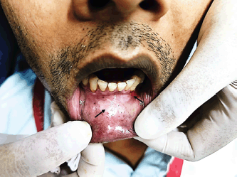
Figure 1. Clinical photograph showing two lesions on lower labial mucosa mimicking leukoplakia.
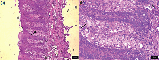
Figure 2. Photomicrograph shows H&E stained section with foamy cells in stroma, (a): low magnification and (b): high magnification.

Figure 3. Photomicrograph shows IHC stained section with foamy cells in stroma positive with CD 68, (a): low magnification and (b): high magnification.
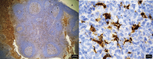
Figure 4. Photomicrograph shows IHC stained section of positive control shows positive staining with CD 68, (a): low magnification and (b): high magnification.
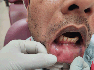
Figure 5. Postoperative follow-up after 6 months.
Discussion
Our case of OVX presents several noteworthy features when compared to the existing literature. While OVX typically manifests as a papillary or verrucoid mass, with the gingiva being the most common site [7], our patient exhibited two smooth-surfaced and Belknap et al [3] white plaques on the lower labial mucosa. This presentation aligns with the findings of Toida and Koizumi [13] and Cebeci et al [14] who reported cases of OVX on the lip, highlighting the clinical diversity of this condition.
The location of our case on the labial mucosa is particularly rare, contrasting with the more common occurrence of masticatory mucosa reported in 70% of cases according to Tamiolakis et al [2]. This atypical presentation posed a diagnostic challenge, initially resembling leukoplakia, which emphasizes the importance of considering OVX in the differential diagnosis of oral white lesions.
Histologically, our case aligned with the characteristic features of OVX, showing aggregates of foamy histiocytes within the connective tissue papillae as mentioned by Barrett et al [6]. The positive CD68 immunohistochemical staining further confirmed the diagnosis, consistent with previous studies Iamaroon and Vickers [9].
Our patient’s history of tobacco use is noteworthy. While OVX is not directly linked to tobacco use, this factor warrants careful consideration, especially given the lesion’s resemblance to leukoplakia. This finding underscores the importance of thorough clinical examination and histopathological analysis in patients with risk factors for oral lesions, as emphasised by Gill et al [15].
Certainly, the patient’s history of tobacco use merits special consideration in the context of this atypical OVX presentation. While OVX is not directly associated with tobacco use, the habit may have contributed to the lesion’s unusual appearance as white plaques, initially mimicking leukoplakia. This underscores the complex interplay between tobacco use and oral mucosal changes, as noted by Reibel [16] in his comprehensive review of tobacco and oral diseases. The presence of tobacco-induced alterations could potentially mask or modify the typical clinical features of OVX, leading to diagnostic challenges. This case highlights the importance of considering a broad differential diagnosis, including both tobacco-related and non-tobacco-related lesions, in patients with a history of tobacco use presenting with atypical oral mucosal changes.
The management of our case through conservative excision aligns with the standard treatment approach for OVX. This is supported by literature reporting low recurrence rates, ranging from 3% Belknap et al [3] to 12% Tamiolakis et al [2] in large case studies.
Interestingly, our patient’s lipid profile showed slightly low VLDL levels, a finding not commonly reported in OVX cases. This observation may warrant further investigation into the potential relationship between lipid metabolism and OVX development.
In conclusion, our case contributes to the growing body of literature on OVX by highlighting its potential to mimic other white lesions, particularly when occurring in atypical locations. It underscores the critical role of histopathological examination in accurate diagnosis and emphasises the need for clinicians to maintain a high index of suspicion for OVX, even when presenting with unusual clinical features.
Conclusion
The presentation of OVX on the labial mucosa as leukoplakia in a patient with a history of tobacco use underscores the need for careful evaluation, accurate diagnosis and appropriate management. While VX itself is benign, it is essential to rule out any co-existing conditions, especially in atypical presentations.
In conclusion, the collective insights from these studies and case reports have enhanced our understanding of OVX, its clinical presentations, potential associations and pathogenesis. OVX remains a rare but diagnostically significant oral lesion. This discussion highlights the importance of continued research in recognising, diagnosing and managing OVX effectively. It also emphasizes the need for a multidisciplinary approach involving clinicians, pathologists and surgeons to provide the best care to affected patients.
Conflicts of interest
The authors declare that they have no conflicts of interest.
Funding
No funding was received for the current case report.
References
1. Shafer WG (1971) Verruciform xanthoma Oral Surg Oral Med Oral Pathol [Internet] 31(6) 784–789 https://doi.org/10.1016/0030-4220(71)90134-4 PMID: 5280461
2. Tamiolakis P, Theofilou VI, and Tosios KI, et al (2018) Oral verruciform xanthoma: report of 13 new cases and review of the literature Med Oral Patol Oral Cir Bucal 23(4) e429–e435 PMID: 29924759 PMCID: 6051686
3. Belknap AN, Islam MN, and Bhattacharyya I, et al (2020) Oral verruciform xanthoma: a series of 212 cases and review of the literature Head Neck Pathol [Internet] 14(3) 742–748 https://doi.org/10.1007/s12105-019-01123-0 PMID: 31898056 PMCID: 7413928
4. Yu CH, Tsai TC, and Wang JT, et al (2007) Oral verruciform xanthoma: a clinicopathologic study of 15 cases J Formos Med Assoc [Internet] 106(2) 141–147 https://doi.org/10.1016/S0929-6646(09)60230-8 PMID: 17339158
5. Santiago E, Rosebush MS, and Owens J, et al (2017) Verruciform Xanthoma: A Retrospective Clinical and Histopathologic Analysis of 90 Cases [Internet] https://spindlerperio.net/wp-ontent/uploads/2018/05/Santiago-2017.pdf Date accessed: 09/03/24
6. Barrett AW, Boyapati RP, and Bisase BS, et al (2019) Verruciform xanthoma of the oral mucosa: a series of eight typical and three anomalous cases Int J Surg Pathol 27(5) 492–498 https://doi.org/10.1177/1066896919827374 PMID: 30727785
7. Mostafa KA, Takata T, and Ogawa I, et al (1993) Verruciform xanthoma of the oral mucosa: a clinicopathological study with immunohistochemical findings relating to pathogenesis Virchows Arch A Pathol Anat Histopathol 423(4) 243–248 https://doi.org/10.1007/BF01606886 PMID: 8236821
8. Nowparast B, Howell FV, and Rick GM (1981) Verruciform xanthoma. A clinicopathologic review and report of fifty-four cases Oral Surg Oral Med Oral Pathol 51(6) 619–625 https://doi.org/10.1016/S0030-4220(81)80012-6 PMID: 6942361
9. Iamaroon A and Vickers RA (1996) Characterization of verruciform xanthoma by in situ hybridization and immunohistochemistry J Oral Pathol Med 25(7) 395–400 https://doi.org/10.1111/j.1600-0714.1996.tb00285.x PMID: 8890055
10. Ishida T, Kawami N, and Kondo S (2006) Verruciform xanthoma on the scrotum Ski Res 5(3) 227–230
11. Hu JA, Li Y, and Li S (2005) Verruciform xanthoma of the oral cavity: clinicopathological study relating to pathogenesis. Report of three cases APMIS 113(9) 629–634 https://doi.org/10.1111/j.1600-0463.2005.apm_238.x PMID: 16218939
12. Rawal SY, Kalmar JR, and Tatakis DN (2007) Verruciform xanthoma: immunohistochemical characterization of xanthoma cell phenotypes J Periodontol 78(3) 504–509 https://doi.org/10.1902/jop.2007.060196 PMID: 17335374
13. Toida M and Koizumi H (1993) Verruciform xanthoma involving the lip: a case report J Oral Maxillofac Surg [Internet] 51(4) 432–434 https://doi.org/10.1016/S0278-2391(10)80363-5 PMID: 8450366
14. Cebeci F, Verim A, and Somay A, et al (2017) Verruciform xanthoma of a lower lip lesion: a new case and review of the literature Case Rep Dermatol 9(2) 130–135 https://doi.org/10.1159/000477961 PMID: 29033816 PMCID: 5624251
15. Gill BJ, Chan AJ, and Hsu S (2003) Verruciform xanthoma Dermatol Online J 20(1)
16. Reibel J (2003) Tobacco and oral diseases: update on the evidence, with recommendations Med Princ Pract 12(SUPPL. 1) 22–32 https://doi.org/10.1159/000069845
17. Huang J, Tseng C, and Tong YJCH, et al (1996) Verruciform xanthoma. Case report and literature review J Periodontol [Internet] 67(2) 162–165 https://doi.org/10.1902/jop.1996.67.2.162 PMID: 8667137
18. Philipsen HP, Reichart PA, and Takata T, et al (2003) Verruciform xanthoma – biological profile of 282 oral lesions based on a literature survey with nine new cases from Japan Oral Oncol 39(4) 325–336 https://doi.org/10.1016/S1368-8375(02)00088-X PMID: 12676251
19. Aihie OP, Azzam MJ, and Haroon A, et al (2022) Verruciform xanthomas in the setting of COVID-19: a case series and review of other conditions associated with this benign cutaneous neoplasm Cureus 14(11) 1–10


