Supporting oncology professionals through education
The content on this site is intended for healthcare professionals only
The content on this site is intended for healthcare professionals only
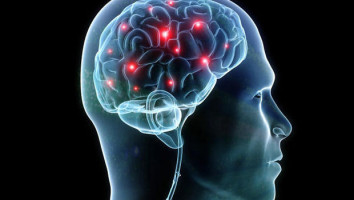
Clinicians from the Johns Hopkins Kimmel Cancer Centre and four other institutions have demonstrated that doctors can gain a wealth of knowledge about a patient’s cancer by using multiple laboratory techniques to study tumour tissue taken from needle biopsies of glioblastoma, a highly aggressive form of brain cancer.
The work, funded by Break Through Cancer and published in the April 28 issue of Nature Communications, has implications for additional cancer types.
Physicians currently limit collection of small tumour samples of glioblastoma because the procedure, stereotactic needle biopsy, requires surgery with a patient sedated with anaesthesia.
Tumour samples are typically taken at the beginning and sometimes at the end of treatment.
But in a new study, researchers injected into the tumour a virus aimed at killing glioblastoma cells.
During the same procedures, surgeons took tumour tissue samples and ran them through multiple types of advanced laboratory techniques, including single-cell RNA sequencing, transcriptomics, metabolomics, proteomics and immune profiling to demonstrate that even small tissue samples can yield additional insights into a tumour’s biology, immune interactions and molecular pathways.
The study found that tissue also could be grafted onto a mouse model for additional analysis.
“One of the major frontiers in oncology is to find better treatments for these tumours, which have limited treatment options. We need a much deeper understanding of why certain treatments work and others don’t work,” explains study co-author Matthias Holdhoff, M.D., Ph.D., co-director of the Brain Cancer Disease Group at the Kimmel Cancer Centre and an associate professor of oncology at the Johns Hopkins University School of Medicine.
During the study, investigators wanted to maximise what they could learn from tissue samples.
“This is a concept that expands beyond just brain cancers,” adds study co-author Chetan Bettegowda, M.D., Ph.D., director of the Metastatic Brain Tumour Centre and medical director of the Ludwig Centre for Cancer Genetics and Therapeutics at the cancer centre.
He also is the incoming director of the Department of Neurosurgery, the Harvey Cushing Professor of Neurosurgery, and a professor of oncology at the Johns Hopkins University School of Medicine.
“Whenever people do needle biopsies, it has been just sufficient to study if the tissue is cancerous, what type and maybe some very simple molecular characterisation. This brings tissue analysis to the modern age. ... Historically [oncologists] haven’t done repeat biopsies because we felt, ‘Oh, what are we going to get that we don’t already know from the original diagnosis?’ It turns out there’s quite a bit to be learned.”
Other participating centres in the study were Memorial Sloan Kettering Cancer Centre in New York City; Dana-Farber Cancer Institute in Boston; MD Anderson Cancer Centre in Houston; and the Koch Institute for Integrative Cancer Research at MIT in Cambridge, Massachusetts.
Source: Johns Hopkins Medicine
The World Cancer Declaration recognises that to make major reductions in premature deaths, innovative education and training opportunities for healthcare workers in all disciplines of cancer control need to improve significantly.
ecancer plays a critical part in improving access to education for medical professionals.
Every day we help doctors, nurses, patients and their advocates to further their knowledge and improve the quality of care. Please make a donation to support our ongoing work.
Thank you for your support.